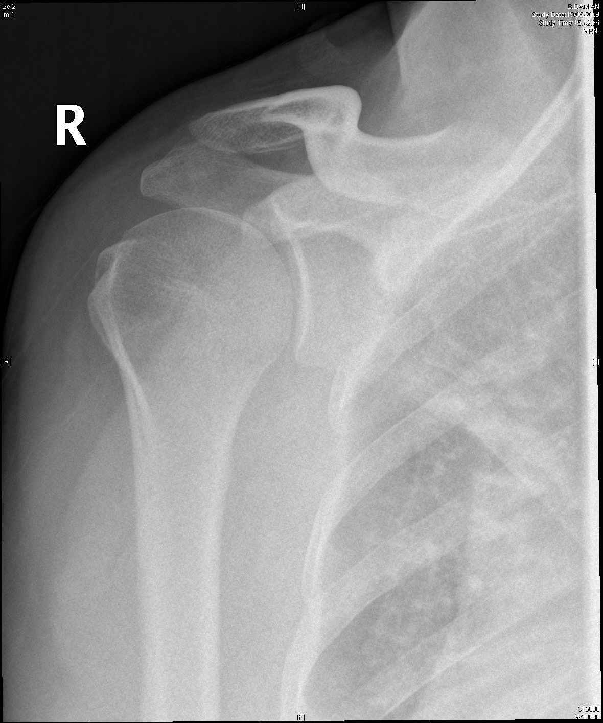
Normal Shoulder
This is done by connecting the humerus ( upper arm) to the scapula (shoulder blade). The scapula in turn is connected to the torso by the clavicle and an array of muscle attaching to the scapula. The genius of this mechanism is that the scapula is also acting like a mobile platform on the chest dramatically increasing the movement of the shoulder.
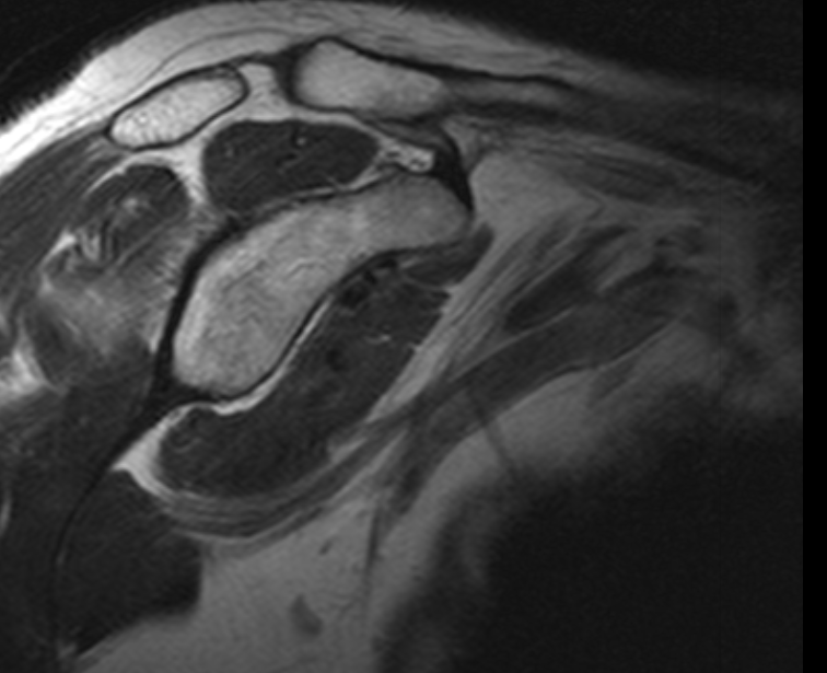
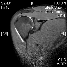
The normal shoulder has several layers.These layers mean that the shoulder joint is several centimeters below the skin. Swelling in the shoulder is not easily seen unless it is very severe.
Swelling in the skin and layer of subcutaneous fat seldom have anything beyond a cosmetic effect on shoulder function (ie lipomas).
The next layer is the deltoid. This muscle is the main source of power in the shoulder and gives the shoulder its rounded contour. Thankfully it is rarely damaged (most common cause of the deltoid not working is an injury to the axillary nerve following a shoulder dislocation).
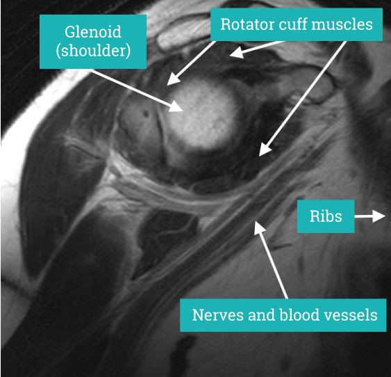
Deep to the deltoid run muscles from the shoulder blade attaching to the top of the humerus just at the edge of the joint. These muscles are known as the rotator cuff. They form a near continuous attachment to the bone so an injury to the tendon (the white sinewy part of the muscle that attaches to the bone) of one muscle often extends into its neighbouring rotator cuff muscle.

There are essentially three groups of muscles in the rotator cuff.
- The muscle at the front of the shoulder (Subscapularis)
- The muscle over the top of the shoulder (Supraspinatus)
- The muscles at the back of the shoulder (Infraspinatus and Teres minor)
The rotator cuff muscles add power and finesse to shoulder movement helping keep the shoulder in position and controlling the finer moments, in particular rotation.
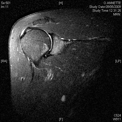
Deep to the rotator cuff lies the deepest layer of soft tissue, the joint capsule. The capsule is a thin but strong fibrous layer that is supple enough to allow full movement but strong enough to stop the shoulder dislocating. It is inflammation in the joint capsule which causes the capsule to stiffen reducing movement causing the symptoms of a frozen shoulder. At the opposite extreme tearing of the capsular attachment to bone is what allows shoulders to dislocate.
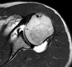
Beneath all these layers lie the cartilage covered bones of the shoulder joint formed by the humeral head (top of the humerus) and the glenoid (the oval joint surface of the scapula). Arthritis in the bones of the shoulder joint results in loss of the white shiny and smooth articular cartilage leaving the much rougher bones directly in contact with each other.
The Nerves and Blood vessels passing down the arm to the hand pass approximately 2 cm away from the joint on its inner surface. Problems with the shoulder seldom affect these structures.
“The normal shoulder has several layers.These layers mean that the shoulder joint is several centimeters below the skin.”
Make an appointment
If you wish to make an appointment to discuss your needs further, please get in touch.
Hermitage Medical Clinic
Old Lucan Road
Dublin 20
Call for appointments: 01 6459562
Telephone: +353 1 645 9000
Fax: +353 1 645 9235
Email: info@johnlunn.ie